Front matter
ICC-3 (028) Interstitial nephritis[L0006–L0011]
L0008
Intended learning objectives[L0013–L0020]
- What
- First learning goal: be able to define interstitial nephritis by the end of the lecture. The definition itself comes later, not on this slide.
- What
- Second goal: be able to list the causes of interstitial nephritis. The causes themselves are not listed here — that comes later.
- What
- Third goal: be able to describe the pathology — what happens inside the kidney tissue. The pathology itself is not described on this slide.
- What
- Fourth goal: be able to summarise the clinical picture, meaning how the disease shows up in a patient. Details are not given here.
- What
- Fifth goal: be able to outline the treatment of interstitial nephritis. The treatment itself is not outlined on this slide.
- What
- Sixth goal: be able to tell the acute and chronic forms of interstitial nephritis apart. The lecture does not yet say how they differ — that's for later slides.
- What
- The line simply reads 'qs' directly under the objectives list, with no further context anywhere in the lecture. What she means by it is not stated.
Outline shown 1 of 9[L0022–L0036]
L0023
L0025
L0027
L0029
Definition[L0038–L0045]
L0039
L0040
L0041
- What
- Tubulointerstitial disease is a broad label for kidney diseases affecting structures other than the glomerulus.
- Where
- Structures in the kidney outside the glomerulus — the filtering unit itself is not the target.
L0043
L0044
- What
- These diseases involve the tubules and/or the interstitium, the tissue around them, while sparing the glomerulus.
- Where
- Tubules and/or interstitium — explicitly not the glomerulus.
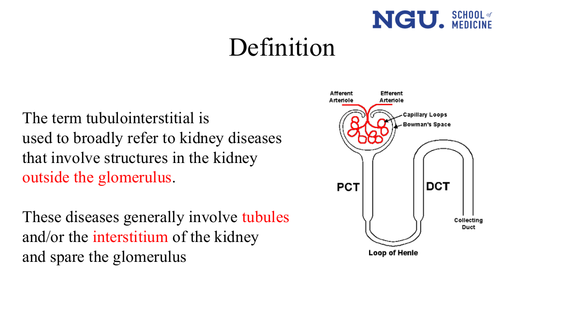
A labelled nephron cross-section. Top: Afferent Arteriole and Efferent Arteriole feeding a glomerular tuft of Capillary Loops inside Bowman's Space. Below it, a tubule runs down as PCT, bends through the Loop of Henle, rises as DCT, and drains into the Collecting Duct.
Draws the exact boundary the text only states in words: arterioles/capillary loops/Bowman's space are the glomerulus; PCT, Loop of Henle, DCT and collecting duct are the tubule — the structures this disease involves while sparing the glomerulus. The diagram also draws the glomerular structures (arterioles, capillary loops) in red against black tubule lines, matching her red highlighting of 'outside the glomerulus', 'tubules' and 'interstitium' in the text beside it.
Pathogenesis[L0047–L0055]
L0048
- What
- Tubulointerstitial disease makes up 10-15% of all kidney disease worldwide.
- Where
- A global figure — she gives a worldwide proportion, not a local one.
- Why
- She gives the global proportion without explaining why tubulointerstitial disease reaches this share of all kidney disease.
L0050
- What
- Damage to kidney cells causes them to display new antigens they did not display before.
- How
- She states injury leads to new antigen expression but not what happens in between.
L0052
L0053
L0054
- What
- Cytokines here come from inflammatory cells (macrophages, lymphocytes) and from the kidney's own cells (proximal tubule, vascular endothelial, interstitial cells, fibroblasts).
- Where
- Both immune-system cells and native kidney cell types are named sources, not one or the other.
- Why
- This cytokine production drives inflammatory cell infiltration into the kidney tissue — the line's own stated downstream effect.
- What
- The disease process described can end in either acute or chronic nephritis — two possible outcomes, not a single fixed course.
- Why
- She states the outcome can be acute or chronic nephritis without saying what determines which one occurs.
Aetiology shown 1 of 2[L0057–L0064]
- What
- Drugs are the single largest cause of tubulointerstitial disease, responsible for 70% of cases.
- Why
- She gives drugs as the leading cause at 70% without explaining why drugs predominate over infection and other causes.
L0059
- What
- One drug-cause category, antibiotics: cephalosporins, ciprofloxacin, erythromycin, penicillin, rifampicin, and sulphonamides are the named examples.
- Where
- One of four drug subcategories listed under the 70% drug total, alongside analgesics, diuretics, and a miscellaneous group.
- What
- A second drug-cause category, analgesics — specifically NSAIDs.
- What
- A third drug-cause category, diuretics — furosemide and thiazides are the two named.
- Where
- One of four drug subcategories under the 70% drug total, alongside antibiotics, analgesics, and a miscellaneous group.
L0063
- What
- A catch-all drug category: allopurinol, carbamazepine, cimetidine, phenytoin, proton pump inhibitors, and valproate.
- Where
- The fourth and catch-all drug subcategory under the 70% drug total, alongside antibiotics, analgesics, and diuretics.
 Full-slide render of the "Drugs (70%)" bullet list, already text at the rows above, plus a small four-pill icon with no labels. Her own emphasis: the section label Drugs (70%): is highlighted yellow, and penicillin, non-steroidal anti-inflammatory drugs, and proton pump inhibitors are independently bolded within the lists — not every drug name is plain as this record previously implied. The icon beside it is decorative.
Aetiology shown 2 of 2[L0066–L0075]
- What
- Infection is the second most common cause, accounting for 15% of cases.
- Why
- How infection produces this disease is stated later: acute pyelonephritis inflames the tubules, producing a neutrophilic cellular infiltrate (Infection causing acute TIN slide).
L0068
- What
- Viral infection is one infectious cause; hantavirus is the one example she names.
- What
- Bacterial infection is the other infectious cause; streptococci is the example she gives.
- What
- In 8% of cases no cause is identified at all.
- What
- TINU — tubulointerstitial nephritis together with uveitis — accounts for 5% of cases.
- Why
- She names TINU as a distinct 5% cause and does not explain further.
- What
- A further cause category, systemic inflammatory disorders, introduced here before its two named examples.
- Where
- A further aetiology category, sibling to drugs, infection, idiopathic, and TINU, introduced before its two named examples.
- What
- Lupus (SLE) is one systemic inflammatory disorder causing this disease, at 2% of cases.
- Why
- She gives lupus as a 2% cause without explaining why or how SLE produces tubulointerstitial injury.
- What
- IgG4-related disorder is the second named systemic-inflammatory cause; she gives no percentage for it.
- Where
- The second named example under systemic inflammatory disorders, alongside lupus — she gives no percentage for it.
Outline exception shown 2 of 9[L0077–L0090]
Not a repeat. The opening Outline listed Pathogenesis as item 2; this one drops it and renumbers everything below, so what was item 3 is now item 2.
- What
- This Outline slide repeats the four-item contents list but without Pathogenesis, which the opening Outline (the row above) listed as item 2.
- Why
- Confirms Pathogenesis was taught, not skipped: she covers it at the rows above, then drops it from every later outline slide.
Why this matters: Shows the lecturer dropped Pathogenesis from the outline after it was taught (she covers it at the rows above, thirty lines before this slide) — its absence here is her own edit, not a gap in these notes.
L0078
L0080
L0082
Drug induced acute inerstitial nephritis[L0093–L0093]
Pathology shown 1 of 2[L0095–L0103]
L0096
L0097
- What
- The kidney tissue itself shows cells packed into the interstitium, often including eosinophils, plus some degree of tubular cell death that varies in severity.
- Where
- In the interstitium between the tubules, and in the tubules themselves.
- How
- She names the infiltrating cells and the tubular necrosis. She does not state how the causative drug injury produces this specific pattern.
L0099
L0100
L0101
L0102
- What
- In rare cases, NSAIDs can also cause a separate glomerular minimal-change lesion alongside the interstitial nephritis, and this second lesion shows up clinically as nephrotic syndrome.
- Where
- In the glomerulus — a different compartment from the tubulointerstitial injury this lecture is otherwise about.
- How
- She states NSAIDs can cause this lesion in addition to TIN. She does not explain the mechanism connecting the drug to the glomerular change.
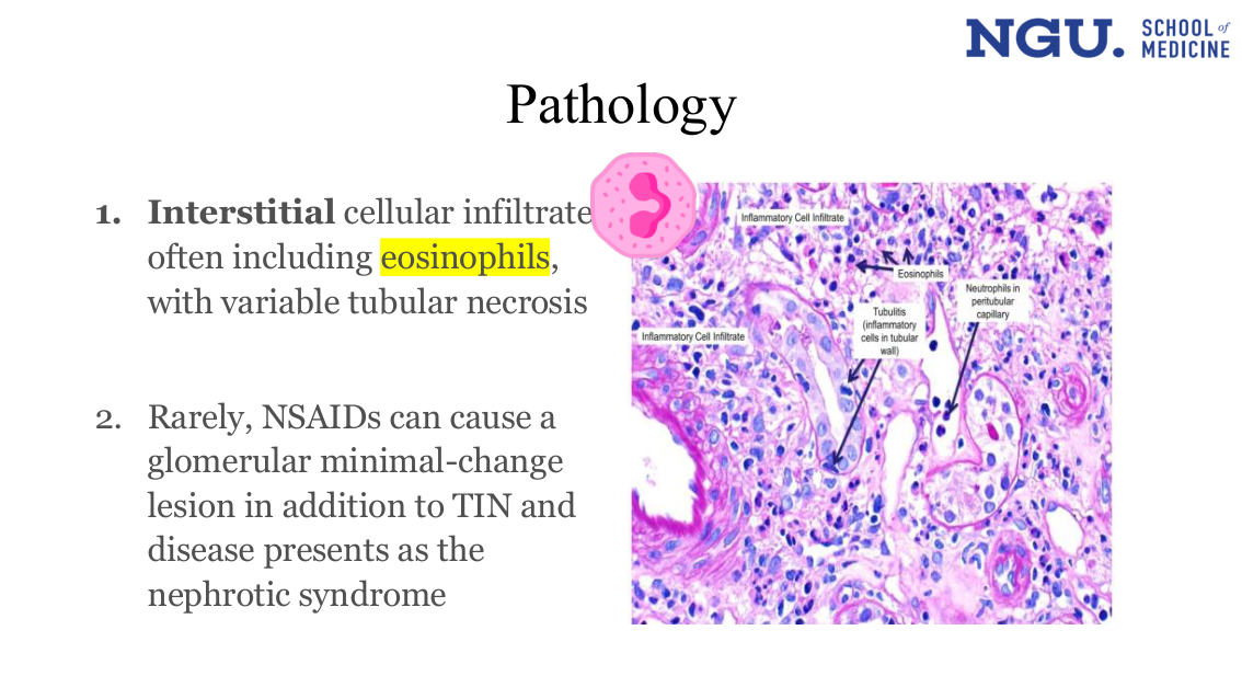
A stained kidney biopsy micrograph with arrows to four labelled findings: Inflammatory Cell Infiltrate (two separate patches of interstitium), Eosinophils, Tubulitis ("inflammatory cells in tubular wall") pointing into a tubule lumen, and Neutrophils in peritubular capillary pointing to a capillary. A decorative cartoon eosinophil icon sits top-left.
Names two findings the text bullet never states: tubulitis (inflammatory cells inside the tubular wall itself) and neutrophils specifically in the peritubular capillary — the text only says "interstitial cellular infiltrate often including eosinophils, with variable tubular necrosis." Her own emphasis, separately: Interstitial is bolded and eosinophils is highlighted yellow in the bullet text itself — the conversion keeps the words and drops both.
Clinical picture of drug induced AIN[L0105–L0113]
- What
- Drug-induced interstitial nephritis can cause no symptoms at all — the only sign is impaired renal function turning up incidentally on a test.
- Where
- The mildest end of this list of presentations — the first entry, ahead of the fever, rash, oliguria, and lab abnormalities that follow.
- What
- Fever, joint pain (arthralgia), and skin rashes can appear together in drug-induced AIN.
- Why
- The lecture calls this condition 'allergic' interstitial nephritis in the investigations section and again in the management flowchart; she never says this triad is what earns the name.
- What
- The kidney injury can reduce urine output (oliguric) or leave it roughly normal (non-oliguric) — both patterns occur.
- Why
- She lists both output patterns as occurring but does not state why the urine-volume response differs from case to case.
- When
- The injury is acute, not chronic.
L0109
- What
- The tubules can malfunction in specific ways: amino acids leak into the urine, glucose leaks into the urine, and renal tubular acidosis occurs.
- Where
- In the tubules themselves — a different site from the rare glomerular lesion above, and a different finding from the interstitial infiltrate that defines this disease.
- How
- She names three specific defects but does not state which tubule segment or mechanism produces each one.
- What
- Many patients with drug-induced AIN also show raised eosinophils in the blood (eosinophilia) and in the urine (eosinophiluria).
- Why
- The lecture calls this condition 'allergic' interstitial nephritis in the investigations section and again in the management flowchart; she never says these two findings are what earn the name.
L0112
- What
- Urinalysis often shows mild protein, blood, and pus cells (white cells) in the urine.
- Why
- She reports the proteinuria as mild here. The rare NSAID glomerular lesion on an earlier slide is the one she says presents as nephrotic syndrome; she does not state the link between the two.
 Full-slide render of the "Clinical picture of drug induced AIN" bullet list, already text at the rows above, plus three unlabelled icons: a cartoon eosinophil, a blood-collection tube, and a urine specimen cup. Her own emphasis: the word Mild is printed in red on the slide, the only coloured word in the clinical picture. The icons beside the bullets are decorative.
Clinical features[L0116–L0116]
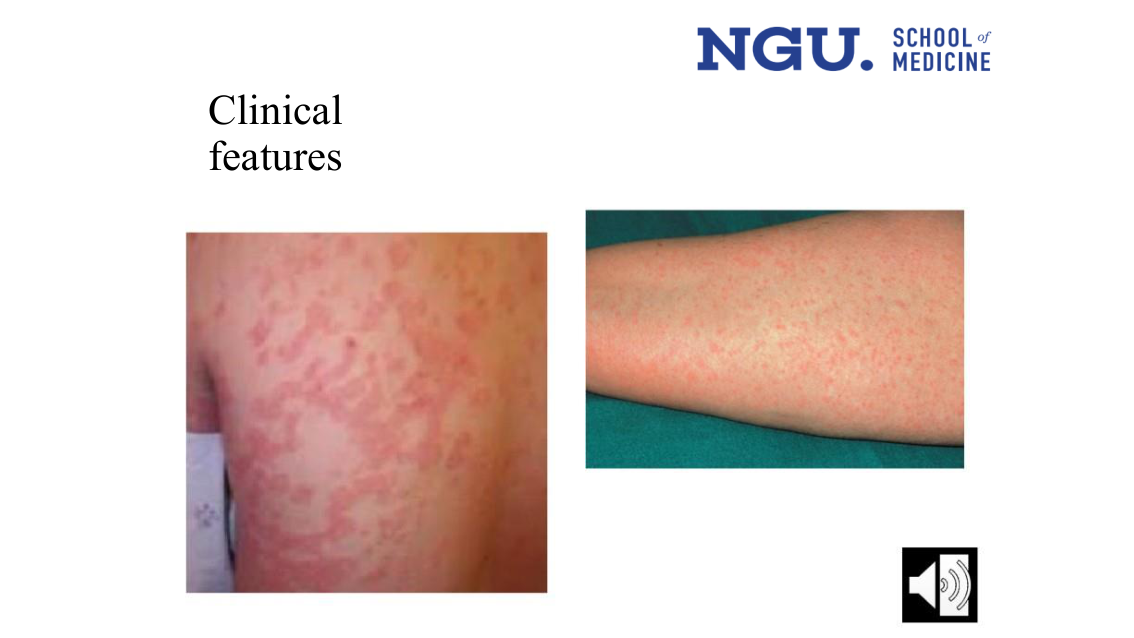
Two clinical photographs, no labels or arrows. Left: close-up of an upper arm/shoulder with a widespread, confluent, blanching erythematous maculopapular (morbilliform) rash. Right: a forearm on a green surgical drape showing a diffuse, finer erythematous maculopapular rash over the skin.
This slide has no bullet text of its own — the heading "Clinical features" carries nothing else. It is the only place in the deck that shows what the drug rash actually looks like: a confluent, blanching, morbilliform eruption, illustrating the "fever, arthralgia, skin rashes" bullet from the earlier Clinical picture slide (the row above), which only names the finding.
Investigations[L0118–L0126]
L0119
L0120
- What
- A kidney that looks normal-sized on ultrasound points toward, but doesn't confirm, a diagnosis of acute kidney disease — the scan alone can't prove it.
L0122
L0123
- What
- Kidney biopsy is the test that actually confirms acute allergic interstitial nephritis — the definitive diagnostic tool, not ultrasound or clinical judgement alone.
- When
- Especially valuable when the clinical picture alone doesn't make the diagnosis obvious.
L0125
- What
- Biopsy shows mononuclear cells, often with eosinophils, infiltrating the kidney — the inflammatory pattern of interstitial nephritis.
- Where
- The infiltrate sits in the renal parenchyma (the interstitium); the glomeruli themselves are spared.
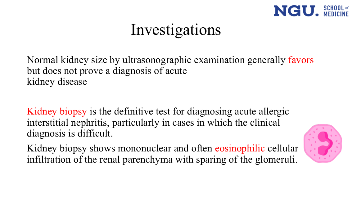 Full-slide render of the Investigations text already at the rows above (ultrasound favors-but-doesn't-prove; biopsy is definitive; biopsy shows mononuclear/eosinophilic infiltrate sparing glomeruli), plus an unlabelled cartoon eosinophil icon. Her own emphasis: three phrases are highlighted red on the slide — favors, Kidney biopsy (the definitive test), and eosinophilic. The text conversion keeps the words and drops the emphasis; the eosinophil icon is decorative.
Outline shown 3 of 9[L0128–L0141]
L0129
L0131
L0133
Treatment[L0143–L0169]
- What
- The first treatment step is simply stopping whichever drug is causing the reaction.
L0145
- What
- High-dose prednisolone, 60 mg daily, may lower the chance of needing dialysis or speed up recovery — an added benefit, not a guarantee ('may').
L0147
L0148
- What
- Most patients regain normal kidney function, but a minority are left with lasting scarring (interstitial fibrosis) and progress to chronic kidney disease.
- Why
- She states some patients are left with fibrosis and CKD. She does not say why those patients, rather than others, progress that way.
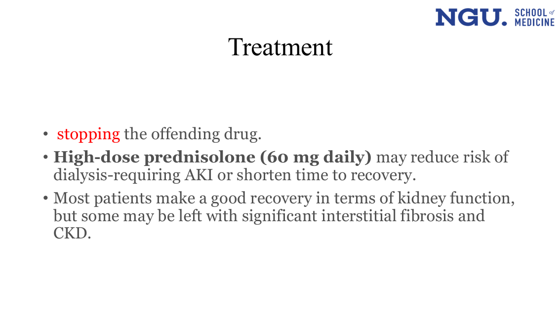 Full-slide render of the Treatment bullet list already text at the rows above (stop the drug; high-dose prednisolone 60 mg daily; most recover, some progress to fibrosis/CKD). No image, icon, or diagram beyond the text. Her own emphasis: stopping is highlighted red, and High-dose prednisolone (60 mg daily) is bolded, singling out the drug and dose. The text conversion keeps the words and drops the emphasis.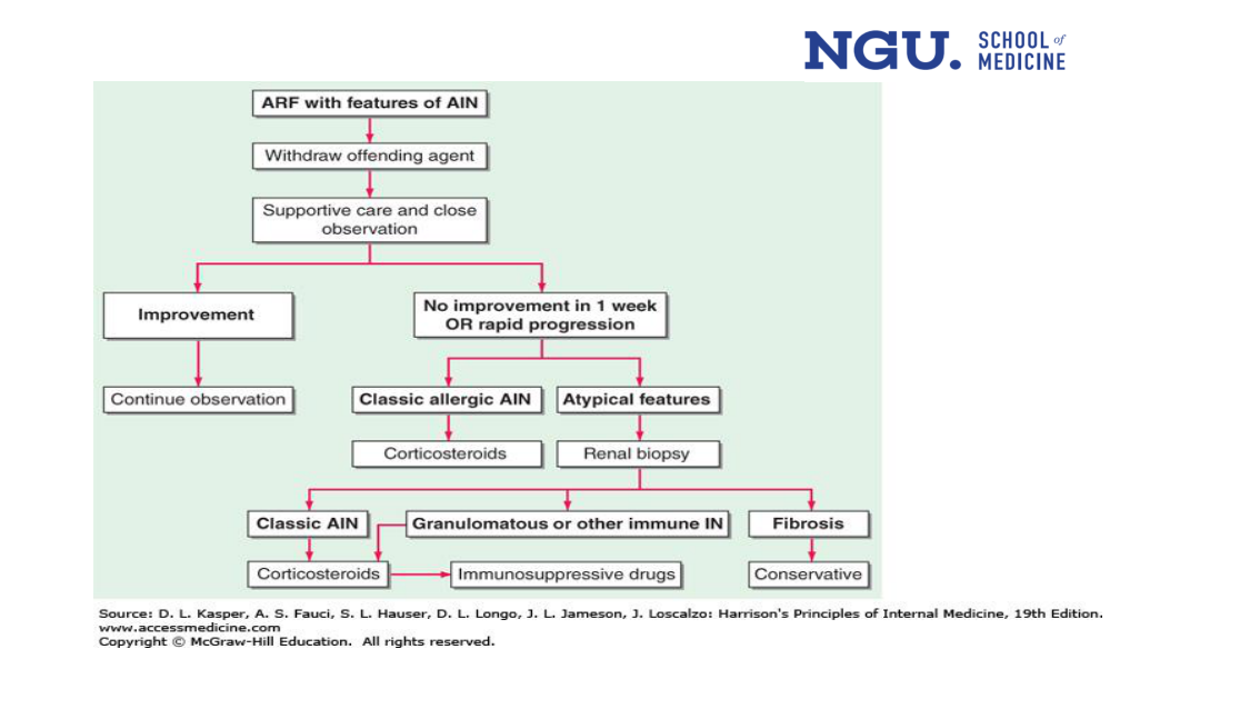
A top-to-bottom flowchart. 'ARF with features of AIN' arrows down to 'Withdraw offending agent', then to 'Supportive care and close observation', which forks into 'Improvement' (arrowing down to 'Continue observation') and 'No improvement in 1 week OR rapid progression'. That box forks again into 'Classic allergic AIN' (down to a 'Corticosteroids' box) and 'Atypical features' (down to 'Renal biopsy'). 'Renal biopsy' alone forks a third time into 'Classic AIN', 'Granulomatous or other immune IN', and 'Fibrosis'. 'Classic AIN' arrows down to a second 'Corticosteroids' box; 'Granulomatous or other immune IN' runs left then down into that same 'Corticosteroids' box, not into 'Immunosuppressive drugs'; 'Fibrosis' arrows down to 'Conservative'. A separate horizontal arrow runs from Corticosteroids to Immunosuppressive drugs, linking the two drug boxes directly.
The arrows show exactly which finding leads to which treatment: both a classic clinical picture and a classic-AIN biopsy result lead to corticosteroids, and the granulomatous/immune biopsy result also arrows into corticosteroids rather than straight to immunosuppressants — that box is reached only via the further, separate arrow running from corticosteroids itself, a link no extracted text line states.
- What
- The algorithm's starting point: acute renal failure whose clinical picture also fits allergic interstitial nephritis (AIN).
- Where
- Sits at the top of the flowchart, above every drug and test decision — the diagnosis that starts the whole management sequence.
This traces her management flowchart (slide 17): each row is the next box in her diagram, not a cause of the one before it.
- What
- The flowchart's fixed opening: diagnosis, then withdraw the drug, then start supportive care, before any branch point.
- Why
- Every case in this algorithm passes through here first; it is the one step no later branch skips.
Why this matters: Every case in this algorithm passes through here first — stopping the drug and starting supportive care come before any branch point, so it is the one step no later branch skips.
- What
- The first action once AIN is suspected: stop the drug or agent causing the allergic reaction.
- Why
- She lists withdrawing the agent as the first step but doesn't state why removing it is expected to help the kidney.
- When
- Done immediately, before supportive care begins and before checking whether the kidney recovers.
- What
- General supportive care plus close watching for whether kidney function returns.
- When
- Follows immediately after the offending drug is withdrawn, and continues until the improve/no-improve branch point is reached.
Same flowchart, its first branch: each row below is what she draws following the finding beside it, not what causes it.
- What
- Supportive care forks two ways: improvement leads to continued observation; no improvement in 1 week, or rapid progression, continues toward treatment.
- Why
- Reaching the improvement lane ends the workup with no biopsy or drug; the other lane is read as classic and reaches steroids without a biopsy.
Why this matters: Marks the fork after supportive care: improving ends the workup with no biopsy or drug, while non-improvement is the branch that reaches steroids without a biopsy, if the picture is read as classic rather than atypical.
- What
- The branch taken when kidney function improves on supportive care alone, without any drug or biopsy.
- When
- Assessed after the withdrawal-and-supportive-care period, before any decision about drugs or biopsy is made.
- What
- If the patient improves, the only further step is ongoing observation — no drug or biopsy is added.
- What
- The trigger for moving past plain observation: either no improvement after a week, or the kidney worsening quickly.
- Why
- She sets a one-week cutoff for judging non-improvement but doesn't state why one week rather than a shorter or longer interval.
- When
- Checked at the one-week mark, or sooner if the patient deteriorates fast.
Same flowchart, the other branch from the same decision point: an atypical picture goes to biopsy instead of straight to treatment.
- What
- From the same non-improvement point, an atypical picture is sent to renal biopsy rather than treated as classic AIN.
- Why
- The mirror-image branch to the classic one beside it — atypical presentation is the one path this flowchart sends to biopsy before treatment.
Why this matters: The mirror-image branch to the classic one beside it — atypical presentation is the one path the algorithm sends to biopsy before any treatment.
- What
- One of two branches once observation has failed: the clinical picture is a textbook allergic AIN.
- When
- Reached only after supportive care has failed to improve the patient, or the patient has worsened quickly.
- What
- The treatment given for a classic allergic AIN presentation.
- What
- The other branch when supportive care fails: the presentation does not fit classic allergic AIN.
- When
- Reached at the same decision point as the classic branch — after failed observation — when the picture is not textbook.
- What
- The test the algorithm calls for when the features are atypical.
- Why
- She orders a biopsy for atypical presentations but doesn't state why atypical features specifically require tissue diagnosis rather than empiric treatment.
- When
- Ordered before treatment is chosen, in place of going straight to steroids.
Same flowchart, its final branch: the biopsy reveals one of three patterns, each drawn to its own treatment box — the biopsy reveals which pattern is present, it does not cause any of them.
- What
- The biopsy shows one of three patterns — classic AIN, granulomatous/immune IN, or fibrosis — each drawn to a different management box.
- Why
- All three biopsy patterns first reach Corticosteroids on this diagram; a separate arrow then runs from Corticosteroids to Immunosuppressive drugs, and fibrosis is the one result that moves to conservative management instead of a drug.
Why this matters: Shows biopsy revealing, not causing, which of three patterns is present: classic AIN and granulomatous/immune IN both arrow into the same Corticosteroids box as the clinical diagnosis, a further arrow from Corticosteroids reaches Immunosuppressive drugs, and fibrosis is the one result past which the algorithm turns conservative.
- What
- One of three biopsy outcomes: the tissue shows a classic allergic AIN pattern.
- When
- Identified only after atypical features prompted a renal biopsy — never reached from the classic clinical branch.
- What
- The treatment for a biopsy-confirmed classic AIN, the same drug used for the clinical-diagnosis branch.
- What
- A second biopsy outcome: a granulomatous or other immune-mediated interstitial nephritis pattern, distinct from classic AIN.
- When
- Identified at the same post-biopsy decision point as the classic and fibrosis outcomes.
- What
- The drug box the diagram links to directly from Corticosteroids by its own arrow, not a box that Granulomatous or other immune IN points to itself.
- Why
- She draws a direct arrow from Corticosteroids to Immunosuppressive drugs but doesn't state when or why treatment would escalate from one to the other.
- What
- The third biopsy outcome: the kidney tissue has scarred.
- When
- Identified at the same post-biopsy decision point as the other two biopsy outcomes, when damage has already progressed to scarring.
- What
- The management given when the biopsy shows fibrosis: conservative care rather than an immune-directed drug.
- Why
- She moves fibrosis to conservative management instead of a drug but doesn't state why scarred tissue is treated differently from the other two biopsy patterns.
Outline shown 4 of 9[L0171–L0184]
L0172
L0174
L0176
Infection causing acute TIN[L0186–L0194]
L0187
- What
- Acute pyelonephritis (a kidney infection) causes inflammation of the tubules and produces a neutrophilic infiltrate — this is how the infection becomes acute TIN.
- Where
- The tubules, not the glomeruli — the infiltrate she describes is tubular, not glomerular.
- How
- She states pyelonephritis triggers tubular inflammation and a neutrophilic infiltrate. She does not state how the infection reaches or damages the tubules — no route or cellular mechanism is given.
- What
- TIN is not only caused by an infection confined to the kidney — a systemic infection elsewhere in the body can also trigger it.
- How
- The next line does name which infections — CMV, BK, and herpes simplex — though only for immunocompromised transplant recipients; no mechanism of damage is given for any of them.
L0190
L0191
- What
- In immunocompromised patients, three specific viruses — cytomegalovirus, BK polyomavirus, and herpes simplex — can cause acute TIN inside a transplanted kidney.
- Where
- In the renal graft itself, in transplant recipients.
- How
- She names the causal viruses — CMV, BK, and HSV — without stating how they damage the graft; no mechanism is given.
- When
- Specifically in immunocompromised patients such as transplant recipients; not stated for immunocompetent patients.
L0193
- What
- Treatment targets the underlying infection — eradicating it with appropriate antibiotics or antiviral agents.
- Why
- She does not state whether treating the infection reverses the tubular inflammation and infiltrate already present, or only halts further damage.
 A clip-art kidney cell and a clip-art virus sit beside the bullets. No labels, arrows or captions on either icon. Her own emphasis: neutrophilic cellular infiltrate is highlighted yellow, and pyelonephritis, systemic infections, and cytomegalovirus, polyoma (BK) and herpes simplex are independently bolded on the slide — not the one phrase this record previously claimed. The text conversion keeps the words and drops all four emphases.
Other Causes of Acute Interstitial nephritis[L0196–L0201]
- What
- Acute TIN can also appear as one feature of a multisystem inflammatory disease, not only from infection or drugs.
- Why
- She groups TIN as a feature of these diseases without stating why or how multisystem inflammatory disease produces tubulointerstitial injury specifically.
- What
- Sjögren's syndrome is listed as one multisystem disease that can cause TIN as part of its picture.
- What
- SLE (lupus) is another multisystem disease listed here as a cause of TIN.
- What
- Sarcoidosis is the third multisystem disease listed here as a cause of TIN.
- What
- TIN with uveitis syndrome (TINU) is named here as a distinct entity — tubulointerstitial nephritis occurring together with uveitis.
- Where
- Kidney and uveitis together, not the kidney alone.
- Why
- She names the syndrome and does not explain why kidney and uveitis occur together.
Chronic interstitial nephritis shown 1 of 3[L0204–L0204]
Outline shown 5 of 9[L0206–L0221]
This Outline slide repeats the same four-item (the opening copy had five - Pathogenesis was dropped after it) contents list given four times earlier in the lecture (the rows above), but here — for the first time — item 4's sub-list gains two extra points, Pathology and Clinical picture, that none of the earlier copies have.
2 sub-points -> 4 sub-points, first appearing here
- What
- This outline slide is the first of its repeats to add Pathology and Clinical picture under item 4, Chronic Tubulointerstitial nephritis.
- Why
- Marks a real change in her plan, not a rendering repeat — the two new sub-points appear only from this slide onward.
Why this matters: Flags a real change in the lecturer's own plan, not a repeat: this fifth pass through this outline is the first to promise Pathology and Clinical picture under chronic TIN, sections the four earlier copies never listed.
L0207
L0209
L0211
Chronic interstitial nephritis shown 2 of 3[L0223–L0235]
- What
- One of five categories the lecture uses to sort causes of chronic interstitial nephritis: drugs and toxins.
- Where
- Listed first, alongside systemic disease, metabolic, infection and miscellaneous causes.
The lecture sorts causes of chronic interstitial nephritis into five named categories, each with its own examples.
Five categories, nineteen named causes, across two slides.
- What
- Five named categories of chronic-TIN causes, spanning nineteen bullets across two slides.
- Why
- Grouping by category is her own structure for this section, not an inference — each slide heads its bullets with the category name.
Why this matters: This is the lecture's own grouping of the nineteen causes named on these two slides into five categories, which is what lets a reader place any one of them (e.g. sarcoidosis, analgesics) without re-reading both slides bullet by bullet — it does not include the further chronic-TIN causes taught later (obstruction, reflux, recurrent UTI, hypercalcaemia), which sit outside this five-category list.
- What
- Whatever causes ACUTE tubulointerstitial nephritis can also cause the chronic form — analgesics is the example given.
- Why
- Signals this is not a separate list: the acute-disease causes carry straight over into chronic disease.
- How
- The mechanism follows later: analgesic nephropathy causes ischaemic damage to the papillae, which then slough into the calyces (Renal Papillary Necrosis slide).
- What
- Ciclosporin is named as one of five drug/toxin causes of chronic interstitial nephritis in this list.
- How
- She names ciclosporin as a cause and does not state the mechanism of tubulointerstitial damage.
- What
- 5-aminosalicylates is named as one of five drug/toxin causes of chronic interstitial nephritis in this list.
- How
- She names the drug class as a cause and does not state how it injures the interstitium.
- What
- Cadmium, lead and titanium are named together, in one bullet, as causes of chronic interstitial nephritis.
- Where
- Sits in the drugs-and-toxins category, in the same bullet list as ciclosporin, 5-aminosalicylates and irradiation.
- How
- She names the three and does not state how each one damages the interstitium.
- What
- Irradiation is named as a toxic cause of chronic interstitial nephritis.
- How
- She names irradiation as a cause and does not state the mechanism of radiation-induced interstitial damage.
- What
- The second category: systemic diseases that cause chronic interstitial nephritis.
- Where
- Sits alongside drugs/toxins, metabolic, infection and miscellaneous as one of the five named categories.
- What
- Diabetes mellitus is named as a systemic-disease cause of chronic interstitial nephritis.
- How
- She names diabetes as a cause and does not state how it damages the renal interstitium.
- What
- Sickle cell disease, or the carrier trait, is named as a cause of chronic interstitial nephritis.
- How
- She names the sickle-cell state as a cause and does not state how it damages the interstitium.
- What
- Systemic lupus erythematosus or vasculitis is named as a systemic-disease cause of chronic interstitial nephritis.
- How
- She names SLE or vasculitis as a cause and does not state how either damages the interstitium.
- What
- Sarcoidosis is named as a systemic-disease cause of chronic interstitial nephritis; in parentheses she gives its subtype as granulomatous tubulointerstitial nephritis.
- Why
- She names the histological subtype in the parenthetical and does not explain why it impairs kidney function.
- How
- No mechanism is given — the parenthetical names a histological subtype (granulomatous), not how sarcoidosis produces it.
- What
- IgG4-related disease is named as a systemic-disease cause of chronic interstitial nephritis.
- How
- She names IgG4-related disease as a cause and does not state how it damages the interstitium.
Chronic interstitial nephritis shown 3 of 3[L0237–L0249]
- What
- The third category: metabolic causes of chronic interstitial nephritis.
- Where
- Listed on the second slide, before infection and miscellaneous.
- What
- Hyperuricaemia — high blood uric acid — is named as a metabolic cause of chronic interstitial nephritis.
- How
- She names hyperuricaemia as a cause and does not state how urate damages the interstitium.
- What
- Nephrocalcinosis is named as one of three metabolic causes of chronic interstitial nephritis in this list.
- How
- She names nephrocalcinosis as a cause and does not state why it damages the interstitium.
- What
- Hyperoxaluria is named as one of three metabolic causes of chronic interstitial nephritis in this list.
- How
- She names hyperoxaluria as a cause and does not state how it damages the interstitium.
- What
- The fourth category: infectious causes of chronic interstitial nephritis.
- Where
- Listed fourth, after metabolic and before miscellaneous, on the second causes slide.
- What
- HIV is named as an infectious cause of chronic interstitial nephritis.
- How
- She names HIV as a cause and does not state how the virus damages the interstitium.
- What
- Epstein–Barr virus is named as an infectious cause of chronic interstitial nephritis.
- How
- She names Epstein–Barr virus as a cause and does not state how the virus damages the interstitium.
- What
- The fifth, catch-all category for causes of chronic interstitial nephritis that don't fit the other four.
- Why
- She groups hypertension, Balkan and herbal nephropathy and Alport's syndrome together without stating what, if anything, unites them mechanistically.
- What
- Hypertension is named as a miscellaneous cause of chronic interstitial nephritis.
- How
- She names hypertension as a cause and does not state how sustained high pressure damages the interstitium.
- What
- Balkan nephropathy, a regionally-named kidney disease, is listed as a miscellaneous cause.
- Where
- Named for the Balkan region; grouped under 'miscellaneous', not under any other named category.
- Why
- She does not state why chronic interstitial nephritis clusters in the Balkan region specifically.
- What
- Herbal nephropathy — kidney damage from herbal remedies — is listed as a miscellaneous cause.
- How
- She does not name which herbs, or state how they damage the interstitium.
- What
- Alport's syndrome is listed as one of four miscellaneous causes of chronic interstitial nephritis in this list.
- How
- She names Alport's syndrome as a cause and does not state how it damages the interstitium.
The same three bulleted lists as the text (metabolic, infection, miscellaneous), with 'Balkan nephropathy' and 'Herbal nephropathy' highlighted in yellow. No other bullet is highlighted, and no extra causes or values are drawn.
The yellow highlighting singles out Balkan nephropathy and herbal nephropathy specifically — an emphasis no text line states.
Outline shown 6 of 9[L0251–L0265]
Pathology shown 2 of 2[L0267–L0272]
L0268
- What
- A pathology finding in chronic interstitial nephritis: long-standing immune cells infiltrating the tissue.
- Where
- In the interstitium — the tissue around the tubules, not the tubules themselves.
- Why
- What drives it is stated earlier, in Pathogenesis: injury induces new antigens, cytokines from inflammatory and renal cells follow, ending in inflammatory cell infiltration.
- What
- A second finding: the renal tubules waste away.
- Where
- In the tubules, alongside the interstitial infiltration.
- How
- She names tubular atrophy as a pathology finding of chronic TIN. She does not state the mechanism by which the tubules atrophy.
L0271
- What
- A third finding: the interstitium shows widespread swelling or scarring.
- Where
- Spread throughout (generalized) the interstitium, not one patch.
- When
- She lists oedema or fibrosis as alternative interstitial changes. She does not state at what stage of disease each appears.
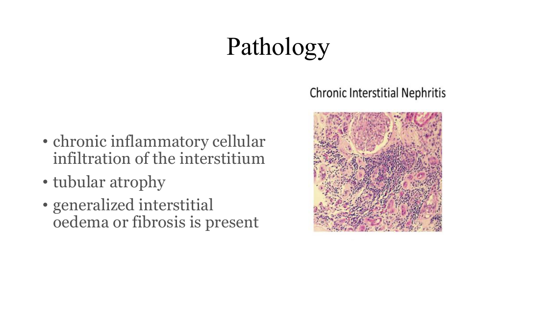
A labelled photomicrograph captioned "Chronic Interstitial Nephritis": a glomerulus at upper left sits amid a dense purple lymphocytic/mononuclear infiltrate filling the interstitium between separated, atrophic-looking tubules, with pale pink patches of fibrosis.
The actual histologic appearance the three bullets only name in words — dense interstitial infiltrate, tubular atrophy, interstitial fibrosis, seen together in one field.
Acute interstitial nephritis[L0274–L0275]
The deck's own sixth and last learning objective is to 'differentiate between acute and chronic interstitial nephritis' (the row above); its outline lists only two headings shared by both sections — Pathology and Clinical picture (the rows above acute, the rows above chronic) — and puts the two names side by side on slide 27 (the rows above).
| Dimension | Acute TIN | Chronic TIN |
|---|---|---|
| Causes | Drugs (70%): antibiotics (cephalosporins, ciprofloxacin, erythromycin, penicillin, rifampicin, sulphonamides), analgesics (NSAIDs), diuretics (furosemide, thiazides), miscellaneous (allopurinol, carbamazepine, cimetidine, phenytoin, proton pump inhibitors, valproate); Infection (15%): viruses e.g. hantavirus, bacteria e.g. streptococci, acute pyelonephritis, and in immunocompromised/transplant patients cytomegalovirus, polyoma (BK) and herpes simplex virus; Idiopathic (8%); TINU (5%) with uveitis; systemic inflammatory disease e.g. SLE (2%), Sjögren, sarcoidosis, IgG4-related disorder | Drugs and toxins, e.g. all causes of acute tubulointerstitial nephritis (analgesics), ciclosporin, 5-aminosalicylates, cadmium/lead/titanium, irradiation; systemic disease e.g. diabetes mellitus, sickle cell disease or trait, SLE/vasculitis, sarcoidosis (granulomatous tubulointerstitial nephritis), IgG4-related disease; metabolic e.g. hyperuricaemia, nephrocalcinosis, hyperoxaluria; infection e.g. HIV, EBV; miscellaneous: hypertension, Balkan nephropathy, herbal nephropathy, Alport’s syndrome |
| Pathology | Interstitial cellular infiltrate, often including eosinophils, with variable tubular necrosis; rarely, NSAIDs can cause a glomerular minimal-change lesion in addition to TIN and disease presents as the nephrotic syndrome | Chronic inflammatory cellular infiltration of the interstitium; tubular atrophy; generalized interstitial oedema or fibrosis is present |
| Clinical picture | Asymptomatic (with impaired renal function found incidentally); fever, arthralgia, skin rashes; acute oliguric or non-oliguric kidney injury; tubular defects as i.e. aminoaciduria, glycosuria, renal tubular acidosis; many have eosinophilia and eosinophiluria; mild proteinuria, hematuria, and the presence of Pus cells | Onset is insidious; patients present with any of polyuria, nocturia; proteinuria (usually slight, <1 g daily); hypertension; uraemia |
| Treatment | Stopping the offending drug; high-dose prednisolone (60 mg daily) may reduce risk of dialysis-requiring AKI or shorten time to recovery; most patients make a good recovery in terms of kidney function, but some may be left with significant interstitial fibrosis and CKD | source silent |
- What
- Both columns share only two of the outline's own headings — Pathology and Clinical picture. Even so the story flips: acute is dramatic and drug-triggered, chronic is silent and structural.
- Why
- That flip is the memory hook — 'acute' means an event you can name and stop; 'chronic' means a state that has already reshaped the tissue.
Why this matters: This is the one place a reader can check pathology, presentation and treatment against each other in one pass instead of across ten slides — the causes cells hold only what's taught by this slide, not her full list; obstruction, reflux, and recurrent UTI, plus hypercalcaemia (her most-common cause), are taught on later slides and aren't in this table. It also surfaces that the lecture never gives chronic TIN a treatment section at all, unlike acute.
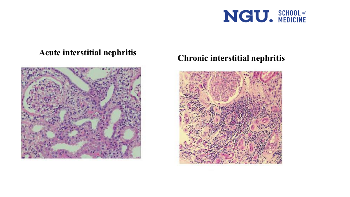
Two labelled photomicrographs. Left, "Acute interstitial nephritis": a glomerulus with a diffuse, dense cellular infiltrate packed between tubules that still look intact. Right, "Chronic interstitial nephritis": a glomerulus with a patchier infiltrate set in paler, more eosinophilic (fibrotic) interstitium around shrunken, atrophic-looking tubules.
The direct visual contrast between the two entities — the heading and caption at the rows above only name them, this image is the entire comparison.
Outline shown 7 of 9[L0277–L0292]
L0278
L0280
L0282
Clinical picture[L0294–L0299]
- Why
- She states the onset is insidious. She does not say why chronic TIN develops gradually rather than acutely.
- When
- The disease starts insidiously — gradually, with no sudden, obvious onset.
- What
- Patients may present with either excessive urination or needing to urinate at night — not necessarily both.
- How
- She lists polyuria and nocturia as presenting features. She does not state why tubular damage produces these urinary symptoms.
- What
- Protein appears in the urine, but usually only mildly — under 1 gram per day.
- Why
- She gives this cutoff, under 1 gram daily. She does not state why chronic TIN keeps proteinuria this mild.
- What
- High blood pressure is another possible presenting feature.
- How
- She lists hypertension as a presenting feature of chronic TIN. She does not state the mechanism by which it raises blood pressure.
- What
- A build-up of waste products in the blood can also be a presenting feature.
- Why
- She lists uraemia as a presenting feature of chronic TIN. She does not state what degree of renal impairment produces it here.
Outline shown 8 of 9[L0301–L0316]
L0302
L0304
L0306
. Chronic Tubulointerstial nephritis[L0318–L0322]
L0320
L0321
- What
- Long-term heavy use of analgesics — historically phenacetin, now mainly NSAIDs — leads to chronic tubulointerstitial nephritis and papillary necrosis.
- How
- The chronic-TIN half is unexplained, but the papillary-necrosis half follows two slides later: ischaemic damage to the papillae, which then slough into the calyces.
Renal Papillary necrosis shown 1 of 2[L0324–L0335]
L0325
L0326
- What
- Ischaemic damage to the renal papillae, which then break off (slough).
- Where
- In the papillae; the sloughed tissue passes into the calyces and out in the urine.
L0329
L0330
L0331
L0332
- What
- A risk factor: long-term use of analgesic combinations — phenacetin with aspirin and caffeine, phenacetin with acetaminophen, or NSAIDs with acetaminophen.
- Why
- These are the specific combinations linked to analgesic nephropathy, the mechanism the previous slide named as causing papillary necrosis.
The previous slide names analgesic use in general as leading to papillary necrosis; this slide gives the exact analgesic combinations that count as the risk factor.
- What
- The general 'analgesic use' cause from the previous slide is spelled out here as three specific drug combinations.
- Why
- Reading the two slides together turns a vague cause into the exact combinations she names as the risk factor.
Why this matters: Reading the two slides together turns a vague 'analgesics cause it' into the specific combinations she actually holds you to.
- What
- Sickle cell disease is listed as a second risk factor for papillary necrosis.
- Why
- She lists sickle cell as a risk factor without stating why it predisposes to papillary necrosis.
- What
- Diabetes is listed as a third risk factor for papillary necrosis.
- Why
- She lists diabetes as a risk factor without stating why it predisposes to papillary necrosis.
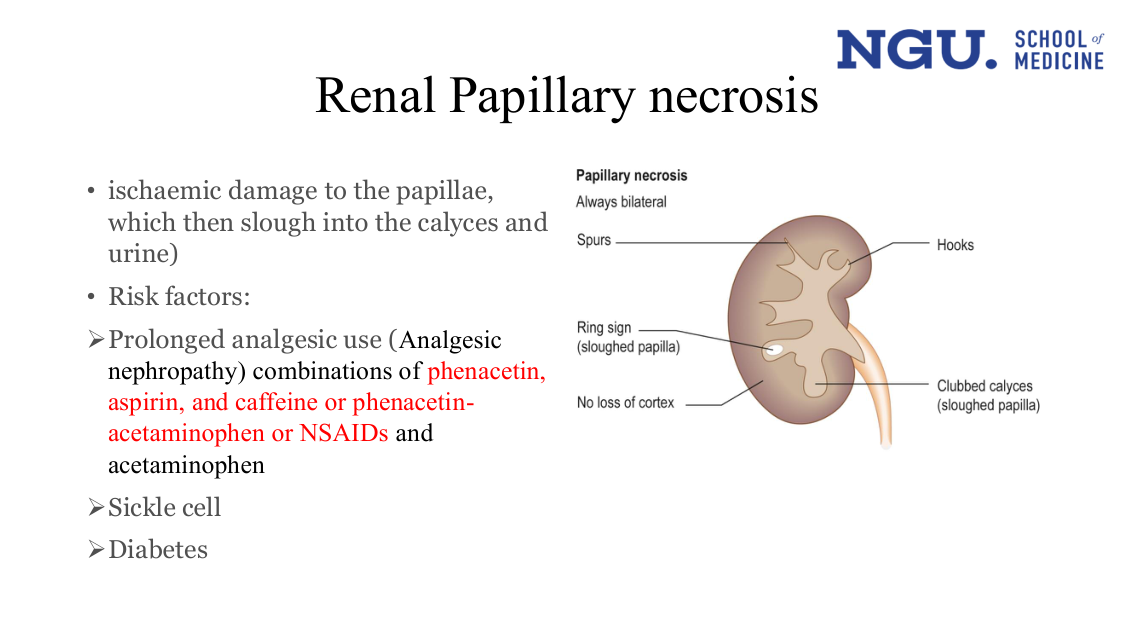
A labelled kidney outline captioned "Papillary necrosis — Always bilateral", with five pointer labels: Spurs and Hooks mark irregular calyceal outlines; Ring sign marks a sloughed papilla sitting free within a calyx; Clubbed calyces (sloughed papilla) marks a calyx left rounded after a papilla detaches; No loss of cortex is labelled on the outer kidney margin.
Every one of these five findings — spurs, hooks, ring sign, clubbed/sloughed-papilla calyces, preserved cortex, and that the process is always bilateral — is stated nowhere in the text rows; the bullets above only give mechanism and risk factors. Her own emphasis, separately: the risk-factor bullet beside the diagram highlights phenacetin, aspirin, and caffeine or phenacetin-acetaminophen or NSAIDs in red — not part of the diagram and not captured in this reading.
Renal Papillary necrosis shown 2 of 2[L0337–L0345]
L0340
- What
- Papillary necrosis can show as blood in the urine, either seen by eye or found only on testing, or as sterile pyuria, pus cells in the urine with no infection.
L0342
L0343
L0344
- What
- Occasionally a sloughed papilla itself causes ureteric colic, or blocks the ureter outright, producing acute obstruction.
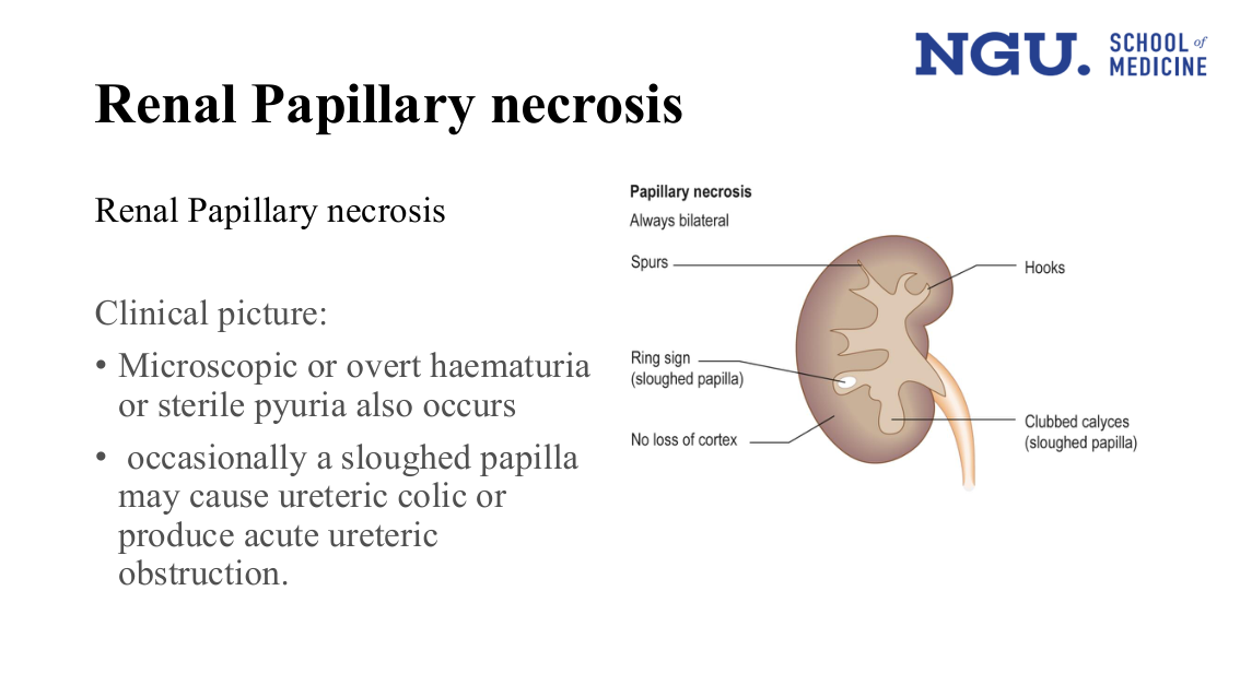
The same labelled kidney diagram as slide32 — captioned "Papillary necrosis — Always bilateral", with Spurs, Hooks, Ring sign (sloughed papilla), Clubbed calyces (sloughed papilla) and No loss of cortex pointer-labelled — now shown beside the clinical-picture bullets (haematuria/sterile pyuria, ureteric colic/obstruction) instead of the risk-factor bullets.
Same five findings as slide32 (spurs, hooks, ring sign, clubbed calyces, preserved cortex, bilaterality), none stated in the clinical-picture text rows above.
Papillary necrosis[L0347–L0347]
Papillary necrosis is given as its own heading a third time (after the rows above), with no new text captured beneath it here.
- What
- A third bare repeat of the same heading, this time introducing a CT image rather than a bullet list.
- Why
- The heading carries no text of its own here; its content is the figure that follows it, not a fourth restatement.
Why this matters: The heading itself is bare, but it sits directly above the slide34 figure record below — the CT image is the content this heading introduces, the radiologic correlate of papillary necrosis that no other slide in the lecture shows.
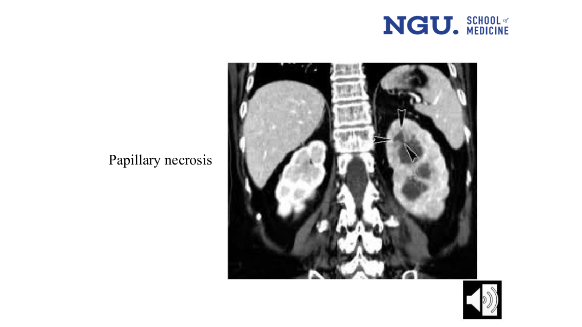
A coronal contrast CT of the abdomen labelled "Papillary necrosis" at left. Two arrowheads point into the left kidney's collecting-system region, marking a filling defect/lucency at the renal papilla consistent with a sloughed papilla; the right kidney shows no such marked defect. A speaker icon (audio) sits at bottom right.
The radiologic (CT) correlate of papillary necrosis — an arrow-marked filling defect in the affected papilla — which no other slide in this lecture shows.
Outline shown 9 of 9[L0349–L0363]
L0350
L0352
L0354
Obstructive Uropathy[L0365–L0378]
L0366
L0367
L0368
- What
- Blocked urine flow, from prostate disease, stones, a tumour, or retroperitoneal fibrosis, can lead to chronic tubulointerstitial disease.
- How
- She states that outflow obstruction causes chronic tubulointerstitial disease. She does not state the mechanism connecting the obstruction to the tubulointerstitial damage.
L0370
- What
- Alongside this, mild protein in the urine and a high-potassium form of renal tubular acidosis are common findings.
L0372
L0373
L0374
- What
- Vesicoureteral reflux, usually present from birth, characteristically leads to focal glomerulosclerosis with nephrotic syndrome and a prominent tubulointerstitial component.
- How
- She states that vesicoureteral reflux results in focal glomerulosclerosis. She does not state the mechanism connecting the reflux to the scarring.
- When
- Usually congenital; the glomerulosclerosis and nephrotic picture are described as showing up in adult life.
L0376
L0377
- What
- Recurrent urinary tract infection can itself produce ammonium magnesium phosphate stones, which further worsen the tubulointerstitial disease and keep the infection going.
- Why
- She states that recurrent UTI causes these stones and that they worsen disease and perpetuate infection. She does not state why infection produces this stone type.
 A plain bullet-text slide with the section title 'Obstructive Uropathy' and four bullets, each already present verbatim in the rows above. No diagram, image or table — only red-highlighted keywords over the same sentences. Her own emphasis: the lead phrase of each bullet is highlighted red on the slide — Obstruction of urinary outflow, Modest proteinuria, Vesicoureteral reflux, and urinary tract infection. The text conversion keeps the words and drops the emphasis.
Types of Obstruction[L0380–L0391]
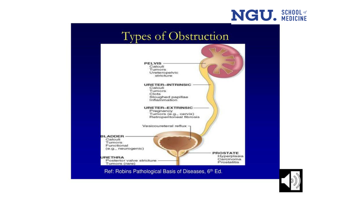
A classification of urinary outflow obstruction by anatomic level, each level labelled with its own causes: Pelvis — calculi, tumours, ureteropelvic stricture; Ureter (intrinsic) — calculi, tumours, clots, sloughed papillae, inflammation; Ureter (extrinsic) — pregnancy, tumours (e.g. cervix), retroperitoneal fibrosis; a separate Vesicoureteral reflux label with no causes given; Bladder — calculi, tumours, functional (e.g. neurogenic); Urethra — posterior valve stricture, tumours (rare); Prostate — hyperplasia, carcinoma, prostatitis. Referenced to Robins Pathological Basis of Diseases, 6th ed.
The labels are organised by anatomic level along the outflow tract, which the flat run of text lines does not itself display as one ordered structure.
- What
- At the level of the renal pelvis, obstruction is caused by stones (calculi), tumours, or a stricture at the ureteropelvic junction.
- Where
- The renal pelvis — the uppermost point in the outflow tract covered by this list.
The urinary outflow tract can be obstructed at any of several levels, and slide 37 gives the typical causes at each.
Six levels, kidney to prostate, each with its own short list of causes.
- What
- Six anatomic levels, pelvis to prostate, each listed with its own short set of causes on slide 37.
- Why
- The levels are her own structure for this slide; several causes (calculi, tumours) repeat across levels rather than being unique to one.
Why this matters: Working out the level of obstruction narrows the cause list, though several causes overlap across levels — calculi appear at three of the six, and tumours at five of six.
- What
- Inside the ureter itself, obstruction can come from stones, tumours, blood clots, sloughed papillae, or inflammation.
- Where
- Intrinsic to the ureter, as opposed to pressure from outside it.
- What
- From outside the ureter, obstruction can come from pregnancy, tumours (e.g. at the cervix), or retroperitoneal fibrosis.
- Where
- Structures outside the ureter compressing it, contrasted directly with the intrinsic causes above.
- What
- Vesicoureteral reflux is listed as its own item in this set of obstruction types, between the ureteric and bladder causes, with no further detail given.
- What
- At the bladder, obstruction is caused by stones, tumours, or a functional cause such as a neurogenic bladder.
- Where
- The bladder, the next level down from the ureters in this list.
- What
- At the urethra, obstruction is caused by posterior valve stricture, or — stated as rare — tumours.
- What
- At the prostate, obstruction is caused by hyperplasia (enlargement), carcinoma, or prostatitis.
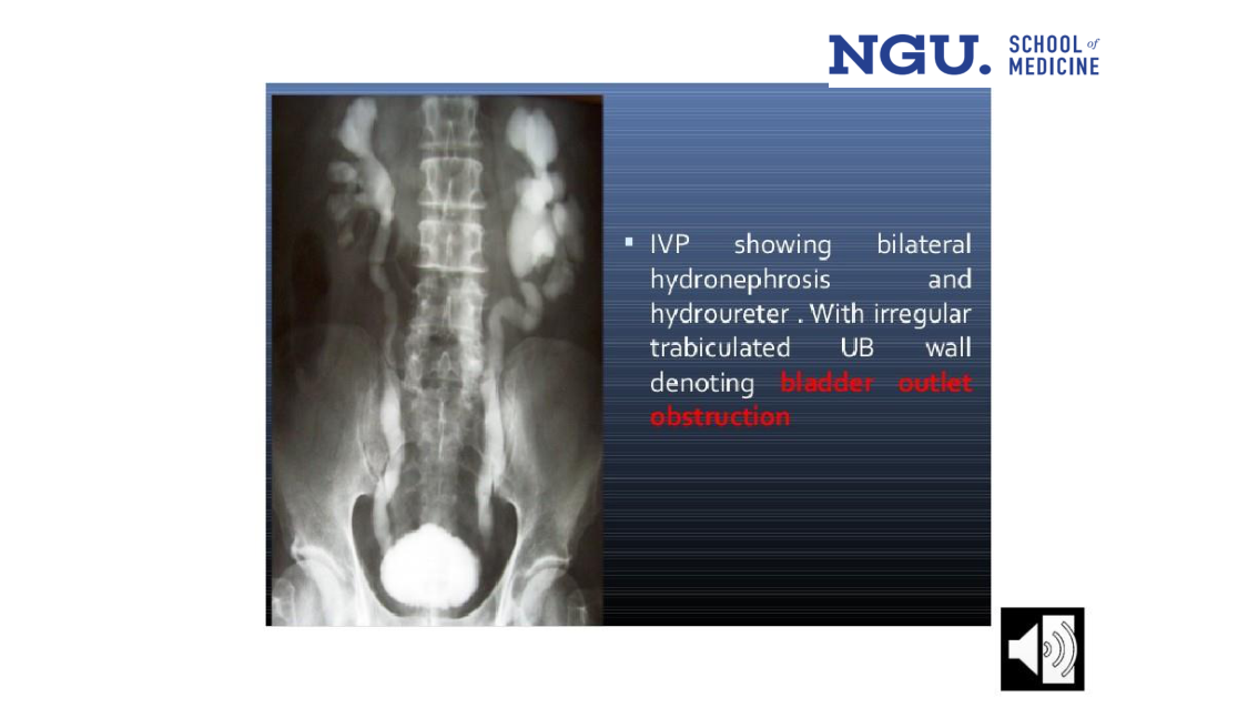
A plain IVP film. Both pelvicalyceal systems are dilated and both ureters are wide down their length; the opacified bladder sits low in the pelvis with an irregular, ragged outline rather than a smooth one.
The film shows the dilatation is bilateral and symmetrical and runs the whole length of both ureters, and the opacified bladder has an irregular, ragged outline — extent the text line names but does not itself draw. Her own emphasis, separately: 'bladder outlet obstruction' is bold and red in the caption text.
- What
- The caption printed beside the film: both collecting systems and both ureters are dilated, and the bladder wall is irregular and trabeculated.
- Where
- At the bladder outlet — the line itself reads 'irregular trabiculated UB wall denoting bladder outlet obstruction.' It does not state why an outlet-level block dilates both collecting systems rather than one.
- Why
- The irregular, trabeculated bladder wall is what the line says denotes bladder outlet obstruction; she does not state why obstruction produces that wall change.
Lead nephropathy[L0393–L0404]
L0394
L0395
- What
- Occupational lead exposure occurs in industrial settings — welders, smelters, battery workers, painters, and restorers of old buildings.
- When
- She names poor ventilation as the setting that raises exposure. She does not state how much lead, or for how long, counts as a 'toxic amount.'
L0397
L0398
- Why
- Without appropriate testing or a careful exposure history, this hypertension gets misdiagnosed as ordinary hypertensive kidney disease rather than recognised as lead-related.
L0400
L0401
L0402
- What
- Lead nephropathy patients tend to have disproportionately worse hyperuricemia than patients with other kidney diseases, and gout is consequently common.
- How
- Stated cause: lead's unique effect on urate metabolism drives the excess hyperuricemia and the resulting gout.
- What
- A restatement of the lead-hyperuricemia link from two lines earlier, here without the urate-metabolism mechanism she gave there.
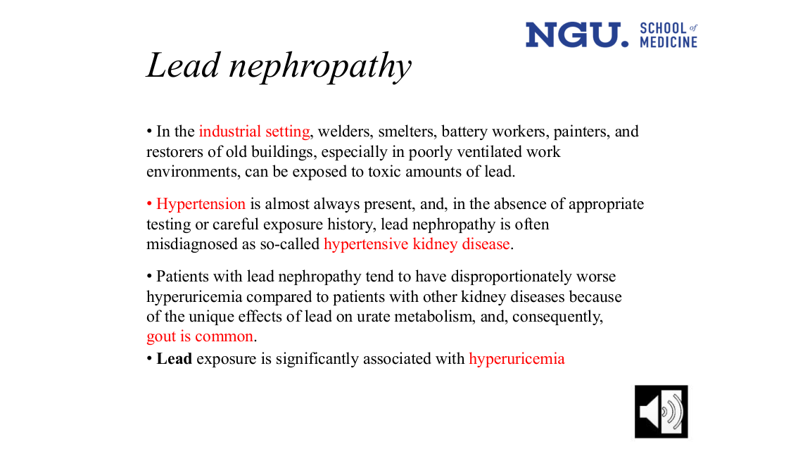 A plain bullet-text slide titled 'Lead nephropathy' with four bullets, each already present verbatim in the rows above (industrial exposure, hypertension misdiagnosed as hypertensive kidney disease, disproportionate hyperuricemia/gout, lead-hyperuricemia association). No diagram, image or table — only red-highlighted keywords over the same sentences. Her own emphasis: five phrases are highlighted red on the slide — industrial setting, Hypertension, hypertensive kidney disease, gout is common, and hyperuricemia — plus Lead bolded in the last bullet. The text conversion keeps the words and drops the emphasis.
Metabolic disorders and nephropathy[L0406–L0419]
- What
- Hypercalcemia is named as the single most common cause in this section on metabolic disorders and nephropathy.
L0408
L0409
L0410
- What
- Chronic hypercalcemia can arise from primary hyperparathyroidism, sarcoidosis, multiple myeloma and other neoplasms (particularly with bone metastases), or vitamin D intoxication.
L0412
L0413
- What
- Unlike the chronic-hypercalcemia causes listed just above, even a transient calcium rise is enough here to leave lasting kidney damage.
- How
- She states transient hypercalcemia causes chronic renal insufficiency limited to the distal tubules. She does not explain how a temporary calcium rise produces permanent tubular injury.
L0415
- What
- Clinically, polyuria and a urine-concentrating defect are common features.
L0417
L0418
- What
- Radiologic exams may show nephrocalcinosis, and renal stone formation can complicate hypercalcemia.
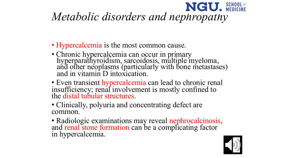 A plain bullet-text slide titled 'Metabolic disorders and nephropathy' with five bullets, each already present verbatim in the rows above (hypercalcemia as most common cause, its chronic causes, transient hypercalcemia and distal tubular injury, polyuria/concentrating defect, nephrocalcinosis/stones on imaging). No diagram, image or table — only red-highlighted keywords over the same sentences. Her own emphasis: five phrases are highlighted red on the slide — Hypercalcemia (bullet 1), hypercalcemia (bullet 3), distal tubular structures, nephrocalcinosis, and renal stone formation. The text conversion keeps the words and drops the emphasis.
Nephrocalcinosis[L0421–L0421]
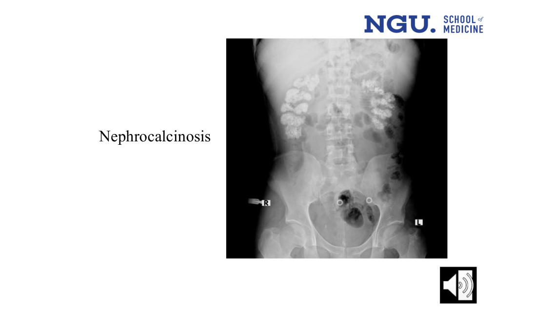
A plain abdominal X-ray (KUB) under the title 'Nephrocalcinosis'. Both kidneys show dense, clustered calcific densities within the renal outlines, more extensive on the right; two round radiopaque ring markers sit near the midline over the pelvis. Side markers 'R' and 'L' orient the film. No other labels or captions.
The actual radiographic pattern of bilateral nephrocalcinosis — clustered renal calcifications on plain film — which no text line in this lecture describes; the surrounding text (the row above, slide 40) only states that imaging 'may reveal nephrocalcinosis' without showing what that looks like.